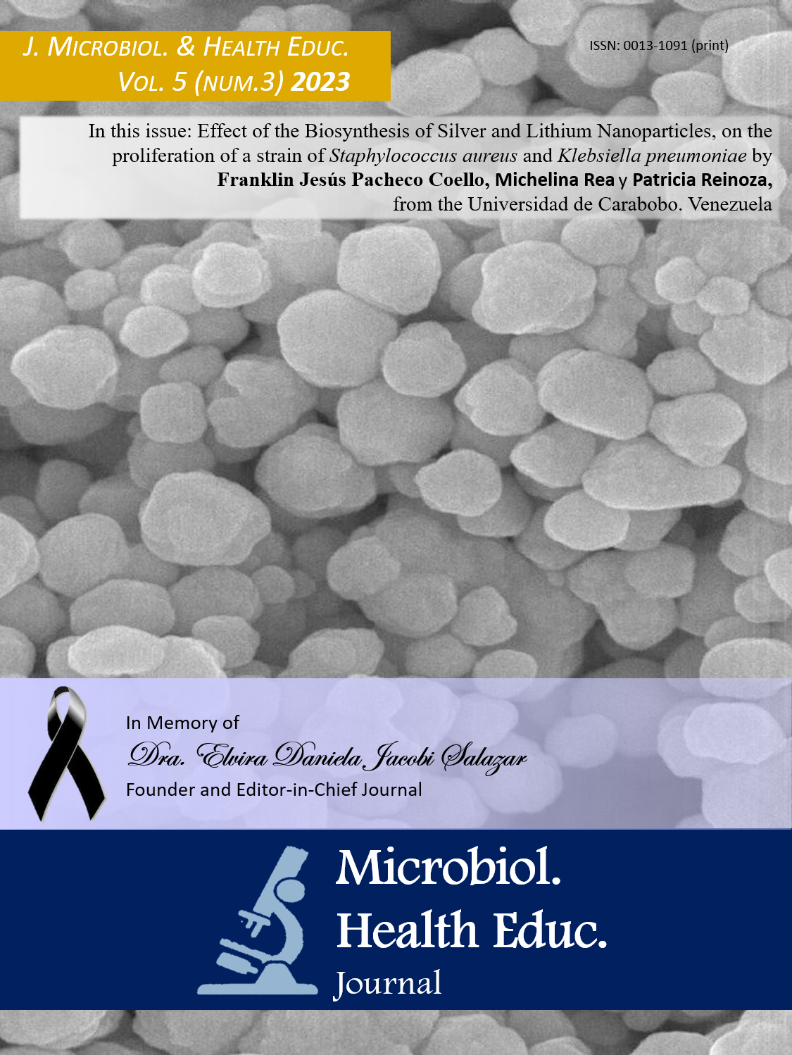
Effect of the Biosynthesis of Silver Nanoparticles (AgNPs) and Lithium (LiNPs), obtained by reduction with Quercetin, on the proliferation of a strain of Staphylococcus aureus (ATCC 25923) and Klebsiella pneumoniae (ATCC 700603)
Keywords:
nanomaterials, morphology, antimicrobial activity, green synthesisAbstract
Introduction: The growing resistance to antibiotics has led to the design and evaluation of new alternatives against various pathogens of clinical interest, including Staphylococcus aureus Klebsiella pneumoniae. Among these alternatives are the so-called metallic nanoparticles, which have exhibited interesting antimicrobial potential. Objective: The study aimed to evaluate the effect of the biosynthesis of silver nanoparticles (AgNPs) and Lithium (LiNPs) on the proliferation of a strain of S. aureus and K. pneumoniae. Methodology: AgNPs and LiNPs were synthesized using lithium and silver salts and quercetin as a reducing agent. These NPs were characterized by scanning electron microscopy (SEM). To evaluate the antimicrobial effect of the NPs, a range of concentrations was established (0.2; 0.4; 0.6; 0.8; and 1 mg/mL). The bacteria were exposed to these concentrations by taking optical density readings every 60 min for 6 h at 600 nm and one at 24 h to calculate the minimum inhibitory concentration (MIC).Results: The AgNPs had a size of 38 nm and 36 nm for the LiNPs, both with spherical morphology and certain aggregations. For all the concentrations evaluated, inhibition of bacterial growth was observed with a minimum inhibitory contraction (MIC) close to 0.8 mg/mL-AgNPs and 0.6 mg/mL-LiNPs against S. aureus and 1 mg/mL- AgNPs and 0.6 mg/mL-LiNPs against K. pneumoniae. Conclusions: The biosynthesis of metallic nanoparticles constitutes a future alternative for the treatment of bacterial infections. AgNPs and LiNPs exhibited excellent performance against S. aureus and K. pneumoniae over the entire range of concentrations.
References
Organización Mundial de la Salud. Resistencia a los antimicrobianos. [On line] 2021 [Citado 30 de abril 2023]. Disponible en: https://www.who.int/es/news-room/fact sheets/detail/antimicrobial-resistance
Tong S, Davis J, Eichenberger E, Holland, T, Fowler Y. Staphylococcus aureus. Infections: epidemiology, pathophysiology, clinical manifestations, and management. Clin Micro Rev, 2015; 28(3): 603-661.
Cueto M, Pascual A. Microbiología y Patogenia de las infecciones producidas por Staphylococcus aureus. En: Pahissa A, Soler H, Soto A, Matos L, Serrano E, Roig MA editores. Infecciones producidas por Staphylococcus aureus. 1a ed. Barcelona (España): Marge Books ed.; 2009. P. 15- 29.
Podschun R, Ullmann U. Klebsiella spp. as nosocomial pathogens: epidemiology, taxonomy, typing methods, and pathogenicity factors. Clin Microbiol Rev. 2018; 11(4):589-603.
Martin RM, Bachman MA. Colonization, infection, and the accessory genome of Klebsiella pneumoniae. Front Cell Infect Microbiol. 2018; 8(2):4-12.
Ramyadevi J, Jeyasubramanian K, Marikani A, Rajakumar G, Rahuman A.A. Synthesis and antimicrobial activity of copper nanoparticles. Materials Letters. 2012; 71(1):114-116. https://doi.org/10.1016/j.matlet.2011.12.055
Hajipour MJ, Fromm KM, Ashkarran A A, Jiménez de Aberasturi D, De Larramendi IR, Rojo T, Serpooshan V, Parak WJ, Mahmoudi M. Antibacterial properties of nanoparticles. Tren biotechol, 2012:30(10), 499-511. https://doi.org/10.1016/j.tibtech.2012.06.004
Albrecht M.A, Evans C.W. Raston C.L. Green chemistry and the health implications of nanoparticles. Green Chem. 2006; 8(5):417-424. https://doi.org/10.1039/b517131h
Kasithevar M, Saravanan M, Prakash P, Kumar H, Ovais M, Barabadi H, Shinwari ZK. Green synthesis of silver nanoparticles using Alysicarpus monilifer leaf extract and its antibacterial activity against MRSA and CoNS isolates in HIV patients. J Interdis Nanomed. 2017; 2(2): 131-141. https://doi.org/10.1002/jin2.26
Sarkar D, Paul G. Green Synthesis of Silver Nanoparticles using Mentha asiatica (Mint) Extract and Evaluation of their Antimicrobial Potential. Int J Cur Res Bio . 2017; 4(1): 77-82. https://doi.org/10.20546/ijcrbp.2017.401.009
Rodríguez-León E, Iñiguez-Palomares R, Navarro R.E, Herrera-Urbina R, Tánori J, Iñiguez-Palomares C, Maldonado A. Synthesis of silver nanoparticles using reducing agents obtained from natural sources (Rumex hymenosepalus extracts), Nano Res Lett. 2013; 8(1): 318. DOI: https://doi.org/10.1186/1556-276X-8-318
Gondwal G.J.N. Pant, Biological evaluation and green synthesis of silver nanoparticles using aqueous extract of Calotropis procera, Int. J. Pharm. Biol. Sci.2013; 4(4): 635-643.
Triana P, De Freitas A, Gómez, K. Estudio de las bacterias que afectan al pie diabético: crecimiento in vitro, efectos de la insulina y susceptibilidad a antibióticos. ´[Tesis de grado en Bioanalisis] Maracay, Venezuela, Universidad de Carabobo, 2008.
Jayandran M, Haneefa MM, Balasubramanian V. Green synthesis of copper nanoparticles using natural reducer and stabilizer and an evaluation of antimicrobial activity. J Chem Pharm Res. 2015;7:251–9
Ramón A.B, Álvarez AM, Cortez-Valadez AR, Britto-Hurtado AL. Bueno O, Flores-Lopez NS , Hernández-Martínez AR. Raman scattering and optical properties of lithium nanoparticles obtained by green synthesis. Vibra Spectro. 2015; 77 (2): 5-9
Khodadadi S, Mahdinezhad N, Fazeli-Nasab B, Heidari MJ, Fakheri B, Miri A. Investigating the Possibility of Green Synthesis of Silver Nanoparticles Using Vaccinium arctostaphlyos Extract and Evaluating Its Antibacterial Properties. BioMed Res Inter. 2021; 1(1): 1-13. https://doi.org : 10.1155/2021/5572252.
Figuera-Calderon M. Síntesis de nanopartículas de hierro a partir del extracto acuoso de hojas de Eucalyptus robusta Sm. y evaluación de su actividad antimicrobiana. [Tesis de grado para Licenciada en Quimica], Caracas, Venezuela, 2019.
Avila-Novoa, M. G., Iñiguez-Moreno, M., González-Gómez, J. P., Zacarías-Castillo, E., Guerrero-Medina, P. J., ... & Gutiérrez-Lomelí, M. (2018). Detection of enterotoxin genes of Staphylococcus aureus isolates from food contact surfaces in the dairy industry of Jalisco, Mexico. Biotecnia, 20(2), 72-78. https://doi.org/10.18633/biotecnia.v20i2.602
Yuan J, Chen C, Cui J, Chan Y, Liung F, Hanh P. Fatty liver disease caused by high-alcohol-producing Klebsiella pneumoniae. Cell Metab. 2019; 30(4):675-688.
Downloads
Additional Files
Published
How to Cite
Issue
Section
License
Copyright (c) 2023 Journal of Microbiology & Health Education

This work is licensed under a Creative Commons Attribution-NonCommercial-NoDerivatives 4.0 International License.


